The Optimax Complete Eye Examination
The Optimax Complete Eye Examination is a lot more than just your typical eye test, it is an eye opening experience in itself, and an amazing way to discover more about your precious eyes.
Not only does this eye exam help to determine eye treatment suitability, it is also a full on eye health screening - where symptoms of eye diseases or ocular disorders such as glaucoma, cataract, diabetic retinopathy, keratoconus, and lazy eyes can be picked up.
The Complete Eye Examination
The eye examination is divided into 6 main sections, namely : Vision, Cornea, External Ocular Health, Muscle Function, Internal Ocular Health, and a personal counselling and Q&A session with you and your family. An eye examination report can also be prepared and provided to you upon request.
The types of tests being performed during The Optimax Complete Eye Examination will normally include:
- Amblyopia/ lazy eye assessment
- Visual acuity
- Refraction – eye power measurement
- Colour vision test
- Eye muscle assessment
- External eye examination
- Corneal topography (corneal mapping)
- Corneal pachymetry (corneal thickness)
- Dry eye assessment
- IOP measurement (intra-ocular pressure)
- Ocular crystalline lens examination
- Fundus eye examination
The tests above, collectively will give us enough information to advise you on:
- Eye health and vision condition
- Treatment suitability – (eg ASA, LASIK, SMILE, ICL, RLE, Ortho-k, RGP)
Special diagnostic tests
Sometimes, certain patients may require (or request for) extra diagnostic tests in addition to those already provided in our Complete Eye Examination. These other diagnostic tests may include one or a combination of the following:
- Visual Field Test (VFT)
- Optical Coherence Tomography (OCT)
- Optical Coherence Tomography Angiography (OCTA) - Sunway branch
- Fundus Photography

A quick look at the 6 main sections:
Vision
Ambloypia/ Lazy Eye assessment
We’ve met many people who grew up thinking they have a lazy eye, when in actual fact, they don’t.
Worried that your child may develop a lazy eye ? - then this assessment will be very meaningful for you ( if they do have a lazy eye, don’t worry, we’re here to help)
Vision and Eye Power assessment
Discover how well your eyes can actually see, and get yourself an accurate eye power measurement.
Did you know?
Every year at Optimax, we come across hundreds of children who wear glasses with powers too high for them.
Ishihara Colour Vision assessment
A favourite among our patients, the Ishihara colour vision assessment is a useful tool for career and education planning.
Did you know?
People with red-green colour deficiency have a harder time telling if their meat is cooked, because of difficulty seeing the different shades of red.
Cornea
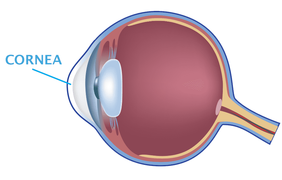
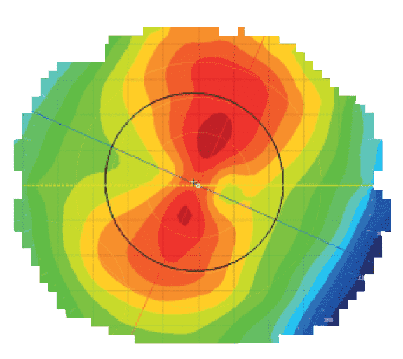
Cornea assessment (health, mapping, and thickness)
When it comes to corneal health assessments, proper checking with a microscope and advanced corneal topography (mapping) is a superb combination.
Did you know?
The measurements from this part, especially cornea thickness is one of the most important components to consider if a person wants to do LASIK.
External ocular Health
In this section, areas on the outer parts of the eye –the conjunctiva, and its surroundings (eyelids, eyelashes), will be examined for signs of pterygiums, papillae/follicles, and other ocular conditions.
*pterygiums – membrane growth on the conjunctiva
*Papillae/follicles – environmental, physiological, or contact lens induced allergic reactions, normally found underneath the eyelids
Eye muscle function
In order to perceive distance accurately and maintain 3D vision, both our eyes need to work together as a team – moving smoothly at the same time, converging and diverging as the need arises.
Eye muscular problems could result in one or a combination of the following:
- double vision (occasionally, or all the time)
- physical appearance of one eye turning out or in (occasionally, or all the time)
- decrease in ability to judge distance, depth, or appreciate 3D vision
- headaches and eye tiredness
- decrease in reading and focusing ability
If you are experiencing any of the above, and can’t seem to figure out why – then this section may be able to provide you with the answers to your questions.
Internal ocular health
The internal parts of the eye generally refer to whatever is inside the eyeball – be it your iris, crystalline lens, retina, optic nerve, or vitreous components.
Inside the eye:
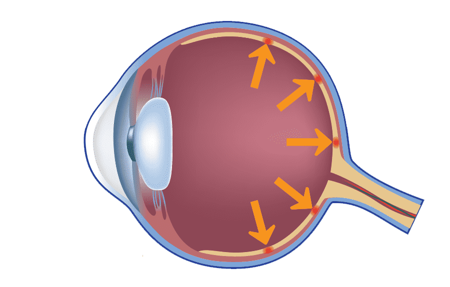
IOP measurement
Imagine your eye as a globe inflated by pressure. Too high of a pressure, and the inner force exerted within the eye can damage your delicate optic nerve, which leads to changes in vision.
Did you know?
The IOP measurement is one of the main components required to accurately diagnose a patient for Glaucoma.
*Glaucoma – a group of ocular disorders that results in optic nerve damage, and in the process reduce an individual’s field of vision. Glaucoma is one of the leading causes of blindness worldwide.
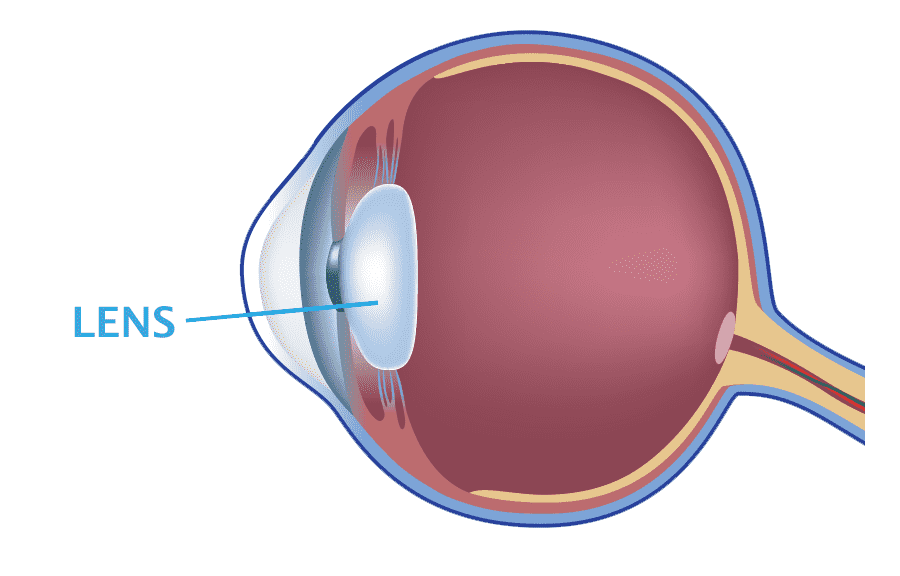
Crystalline Lens inspection
The crystalline lens is a magnificent structure. It allows us to ‘autofocus’, just like an adjustable camera lens. With age, it loses its flexibility and can turn cloudy., thus affecting our ability to see clearly.
Having difficulties with your near reading, or are worried about CATARACTS ? - then this section will be very meaningful for you.
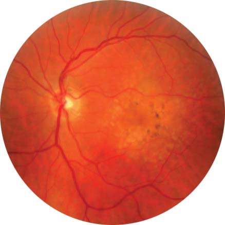
Fundus examination – retina and optic nerve
The retina is an extension of the brain. It converts light into visual signals, which are then transmitted by the optic nerve to be interpreted by the brain!
Did you know?
Just like brain tissue, retina tissue cannot regenerate. Therefore, any potential damage or disorders to the retina must be picked up early to avoid permanent blindness.
Counselling and Q&A session
An Optimax Complete Eye Examination is never complete without a personal counseling and Q&A session with you (and your family, if you feel more comfortable having them around)
The counseling session will cover the following:
- Explanation and run through of your testing results
- Confirmation of treatment suitability, and discussion of your available treatment options
- Explanation on how the treatment is done
- Risks and side effects of your treatment options
- Do’s and don’ts after treatment
- Pricing and payment methods
- Any other questions you may have
Our counseling session is aimed at ensuring that you understand all there is to know about your eyes and treatment options. At Optimax, we will never advise you to undergo a treatment or surgical procedure that you do not fully understand or are not comfortable with.
MAKE AN ENQUIRY OR APPOINTMENT
Talk to us today
Whether you need a simple eye exam, or a complex eye surgery, rest assured that our highly skilled team of ophthalmologists and optometrists will be here to attend to your needs. Call us today, we are here to help!
CONTACT US NOW